
2D Differential Gel Electrophoresis (2D-DIGE) Proteomics & HCP Analysis
ITSIBIO »We offer high-resolution 2D-DIGE gel electrophoresis with LC-MS/MS protein identification to support comparative proteomics, biomarker discovery, and host cell protein (HCP) analysis in biopharmaceutical development.
Reach out to us for more information. Also, click a button below to view our list of citations involving our 2D-DIGE services or to fill out a project submission form to receive a quote. You can read more about the services we offer below.
ITSI-Biosciences delivers an integrated 2D-DIGE and LC-MS/MS proteomics workflow for confident protein expression profiling, biomarker discovery, and characterization of post-translational modifications. Using validated sample preparation kits and protocols, robotic spot picking and in-gel digestion, DeCyder® difference-in-gel analysis software, and advanced mass spectrometry, we generate high-quality, reproducible results from complex biological samples. Your 2D-DIGE study output can include protein identity, pI, molecular weight, relative abundance, and PTM information—enabling downstream biological interpretation and decision-making.
Trusted by NIH programs, biotech and pharmaceutical teams, research institutes, and universities, ITSI-Biosciences brings 15+ years of hands-on expertise in 2D-DIGE, proteomics, and mass spectrometry. We focus on consistent, picture-perfect gels, fast turnaround, and transparent deliverables—backed by standardized methods that reduce gel-to-gel variability. To streamline projects from start to finish, we also offer complementary solutions for protein isolation, protein quantitation, protein qualification, and specialized HCP analysis support, helping organizations move efficiently from sample to actionable data.
What is 2D-DIGE?
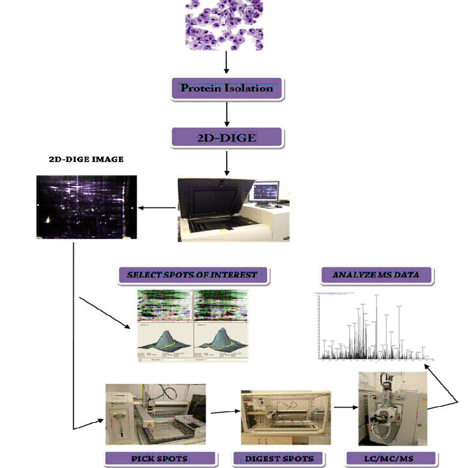
Two-dimensional differential in-gel electrophoresis (2D-DIGE) provides the separation power of classical 2D-PAGE while minimizing gel-to-gel variation by running multiple fluorescently labeled samples in the same gel. Protein samples (commonly labeled with Cy2/Cy3/Cy5 dyes) are combined with a universal internal standard and separated by isoelectric point (1st dimension) and molecular weight (2nd dimension). DeCyder® software then detects, quantifies, and statistically evaluates differentially expressed protein spots for robust comparative and semi-quantitative proteomics.
From gel to identification (2D-DIGE – MS/MS): Differential spots can be excised using automated picking, digested, and identified by LC-MS/MS to confirm protein identity and support pathway-level interpretation. This combined 2D-DIGE and MS/MS approach is widely used in oncology, neuroscience, cardiovascular research, and neurodegenerative disease studies (e.g., Alzheimer’s and Parkinson’s). It can also support workflows targeting post-translationally modified proteins involved in signaling and trafficking, and can be integrated into broader impurity and HCP analysis strategies when characterizing complex biologics-related samples.
2D-DIGE HCP Analysis (Host Cell Protein Profiling)
Key benefits of 2D-DIGE for HCP analysis
- Reduced gel-to-gel variability by multiplexing samples with an internal pooled standard (CyDye labeling).
- Orthogonal separation (pI then molecular weight) to improve resolution of complex HCP mixtures.
- Visualization of proteoforms and PTMs, including charge variants that may be difficult to interpret in 1D-only approaches.
- Quantitative comparison across conditions using DeCyder®-based spot detection, normalization, and statistics.
- MS-ready workflow with excision/in-gel digestion and LC-MS/MS identification of differential spots.
Common applications
- Cell line and clone selection: compare host cell protein expression profiles across candidates.
- Upstream process changes: assess media, feed, and stress impacts on HCP composition.
- Downstream purification monitoring: track HCP clearance across unit operations and identify persistent/co-purifying species.
- Comparability and troubleshooting: highlight HCP shifts between lots, scales, or process conditions.
- Spot-to-protein identification: select differential spots for LC-MS/MS to support targeted risk assessment and process optimization.
2D-DIGE provides a robust, gel-based quantitative proteomics approach for HCP analysis and high-resolution host cell protein profiling in biopharmaceutical development. Samples are labeled with spectrally distinct CyDyes and co-resolved in the same gel alongside an internal pooled standard to enable reliable spot matching and relative quantitation across conditions.
Proteins are resolved by isoelectric point (IEF, 1st dimension) and then by molecular weight (SDS-PAGE, 2nd dimension), delivering orthogonal separation that helps visualize isoforms and post-translational modifications (PTMs)—including charge variants commonly observed in host cell proteins. The internal standard supports consistent spot matching, normalization, and statistical comparisons across large data sets, increasing confidence in differential HCP abundance measurements.
In bioprocess workflows, 2D-DIGE HCP analysis is valuable for monitoring HCP profiles during cell line development, upstream processing, and downstream purification. It supports detection of persistent or co-purifying HCP species, evaluation of process- or stress-induced proteomic shifts, and assessment of clearance efficiency across unit operations. Differential spots can be excised for protein identification by LC-MS/MS, enabling targeted risk assessment and informing process optimization strategies.
As a complementary technique to LC-MS-based proteomics, 2D-DIGE provides strong visualization of proteoforms and reproducible relative quantitation for comparative studies—supporting a broader control strategy for host cell protein characterization across development and manufacturing.
"*" indicates required fields
Citations
Browse through a list of scientific publications and research papers involving our 2D-DIGE Services.
Identification of STAT2 serine 287 as a novel regulatory phosphorylation site in type I interferon-induced cellular responses.
H. C. Steen, S. Nogusa, R. J. Thapa, S.H. Basagoudanavar, A. Gill, S. Merali, C. A. Barrero, S. Balachandran, A. M. Gamero, The Journal of Biological Chemistry
Acute mitochondrial dysfunction after blast exposure: potential role of mitochondrial glutamate oxaloacetate transaminase.
P. Arun, R. Abu-Taleb, S. Oguntayo, Y. Wang, M. Valiyaveettil, J. Long, M.P. Nambiar Journal of Neurotrauma
Modulation of hearing related proteins in the brain and inner ear following repeated blast exposures.
P. Arun, M. Valiyaveettil1, L. Biggemann, Y. Alamneh, Y. Wei, S. Oguntayo, Y. Wang, J.B. Long, M.P. Nambiar1 … Interventional Medicine & Applied Science vol.4 2012 October 1
The effect of selenium enrichment on baker’s yeast proteome.
K. El-Bayoumy, A. Dasa, S. Russell, S. Wolfe, R. Jordan, K. Renganathan, T.P. Loughranb, R. Somiari. Journal of Proteomics-Elsevier Inc. 2011 October 21;.10.1016
Selenium-Responsive Proteins in the Sera of Selenium-Enriched Yeast-Supplemented Healthy American and Caucasian Men.
R. Sinha., I. Sinha., N. Facompreruqui., M. Elmissiry. Journal of Biomedical Science. 2009 September;35(3):559-67.
Proteomics of rat prostate lobes treated with 2-N-Hydroxylamino-1-methyl-6-phenylimidazo [4,5-b] pyridine, 5alpha-dihydrotestosterone, individually and in combination.
Boyiri, T., Somiari, RI., Russell, S., EL-Bayoumy, K. Int. J Oncol. 2009 Sep;35(3):559-67
High-throughput proteomic analysis of human infiltrating ductal carcinoma of the breast.
Somiari RI, Sullivan A, Russell S, Somiari S, Hu H, Jordan R, George A, Katenhusen R, Buchowiecka A, Arciero C, Brzeski H, Hooke J, Shriver C. Proteomics. 2003 Oct;3(10):1863-73.
Albumin depletion method for improved plasma glycoprotein analysis by two-dimensional difference gel electrophoresis.
Brzeski H, Katenhusen R, Sullivan A, Russell S, George A, Somiari RI, Shriver C. Biotechniques 2003 Dec;35(6):1128-32.
Difference In-Gel Electrophoresis in a High-Throughput Environment.
Somiari RI. Russell S, Somiari SB, Sullivan AG, Ellsworth, DL, Brzeski, H, Shriver, CD In Walker J. (Editor) The Proteomics Protocols Handbook. Humana Press Inc., NJ, USA. 2005;pp. 223 – 237.
Proteomics of breast carcinoma.
Somiari RI, Somiari S, Russell S, Shriver CD. J Chromatogr B Analyt Technol Biomed Life Sci. 2005 Feb 5;815(1-2):215-25.
Proteomics in human cancer research.
Somiari RI, Somiari S, Pastwa E, Czyz M. Proteomics. Clinical Applications. 2007 January; DOI: 10.1002/prca.200600369.
