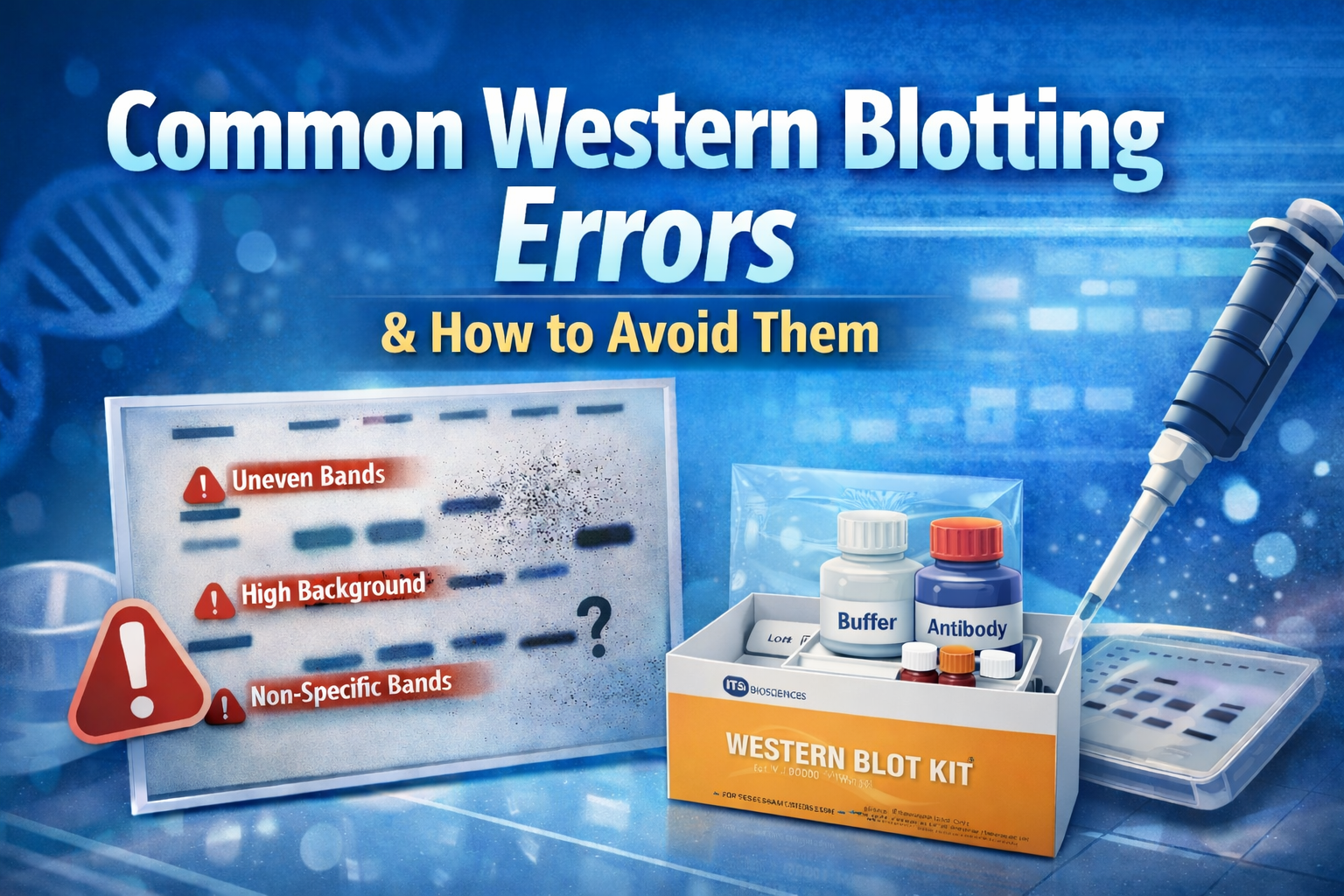
Western blotting is a cornerstone technique in molecular biology, enabling researchers to detect, analyze, and quantify specific proteins within complex samples. However, the multi-step nature of the workflow—sample preparation, electrophoresis, transfer, blocking, antibody incubation, and detection—creates numerous opportunities for error. Even minor inconsistencies can result in weak signals, high background, uneven bands, or misleading data.
Understanding the most common western blotting mistakes and how to prevent them is essential for improving reproducibility, saving valuable lab time, and generating publication-quality results.
1. Poor Sample Preparation
Many western blot failures originate before the gel is ever run. Improper lysis buffers, lack of protease inhibition, or repeated freeze-thaw cycles can degrade proteins and compromise detection.
How to avoid it:
Use freshly prepared lysis buffer supplemented with protease and phosphatase inhibitors when required. Keep samples on ice and minimize handling time to preserve protein integrity. Always quantify protein concentration accurately using assays such as BCA or Bradford to ensure equal loading.
How ITSI-Biosciences Helps:
The ITSI-Biosciences W-Blot™ Western Blot Kit (K-0045) eliminates many early-stage preparation challenges by providing a ready-to-probe western blot containing 20µg of high-quality total protein per lane. By starting with a professionally prepared blot, researchers can reduce variability and focus directly on protein detection.
2. Uneven or Incorrect Protein Loading
Inconsistent band intensity is often caused by unequal protein loading rather than true biological differences. Overloading may create smeared bands, while underloading can produce faint signals.
How to avoid it:
Normalize samples to the same protein concentration and load consistent volumes across lanes. Include a reliable loading control such as β-actin, GAPDH, or tubulin to verify uniform distribution.
How ITSI-Biosciences Helps:
Each W-Blot Kit includes internal standards that enable blot-to-blot and cross-species comparisons, supporting more confident qualitative and semi-quantitative immunoblotting results.
3. Gel Selection and Electrophoresis Errors
Choosing the wrong gel percentage can prevent optimal protein separation. Higher-percentage gels are better for smaller proteins, while lower percentages allow larger proteins to migrate properly. Running gels at excessive voltage can also distort bands.
How to avoid it:
Select gel compositions based on your target protein’s molecular weight and run electrophoresis at recommended voltages. Monitoring the dye front helps prevent overheating and ensures even migration.
Did You Know?
W-Blot™ is generated using high-quality protein samples separated by SDS-PAGE, such as 4–20% gradient gels, ensuring strong resolution before the blot even reaches your lab.
4. Inefficient Protein Transfer
Even perfectly separated proteins are useless if they do not transfer efficiently to the membrane. Incomplete transfer leads to faint bands, while excessive transfer may cause smaller proteins to pass completely through the membrane.
How to avoid it:
Optimize transfer time and voltage according to protein size and membrane type. Nitrocellulose is widely trusted for consistent binding, while PVDF may be preferred for certain applications.
How ITSI-Biosciences Helps:
W-Blot™ arrives on a nitrocellulose membrane that is pre-blocked and ready to probe with your antibody, dramatically reducing hands-on time and transfer-related variability. A PVDF membrane option is also available upon request.
5. Inadequate Blocking
Blocking prevents antibodies from binding nonspecifically to the membrane. Skipping or shortening this step often results in high background noise that can obscure true signals.
How to avoid it:
Block membranes for at least one hour at room temperature or overnight at 4°C using an appropriate blocking agent such as nonfat dry milk or BSA.
How ITSI-Biosciences Helps:
Because each blot is pre-blocked, researchers can bypass one of the most error-prone steps in western blotting and move directly into antibody incubation.
6. Incorrect Antibody Concentrations
Too much antibody increases background, while too little may fail to detect the protein of interest. Unvalidated antibodies frequently produce ambiguous or unexpected bands.
How to avoid it:
Start with manufacturer recommendations but perform dilution optimization experiments whenever possible.
Added Flexibility:
W-Blot™ supports both radioactive and non-radioactive detection methods, allowing laboratories to tailor protocols to their experimental needs.
7. Insufficient Washing Steps
Inadequate washing leaves excess antibody on the membrane, contributing to nonspecific binding and blotchy backgrounds.
How to avoid it:
Perform multiple washes with TBST or PBST while maintaining gentle agitation.
8. Detection and Imaging Problems
Overexposure can saturate strong bands, whereas underexposure may hide low-abundance proteins.
How to avoid it:
Capture multiple exposure times to identify the optimal signal range and use fresh detection reagents.
Pro Tip:
Because W-Blot can be re-probed multiple times, researchers can evaluate additional targets without preparing a new blot—saving both samples and valuable research time.
Reduce Troubleshooting with a Ready-to-Probe Western Blot
Our Western Blot Kit (K-0045) is designed to remove many of the technical barriers associated with traditional western blot workflows.
This ready-to-use blot supports protein analysis from human, animal, or plant tissues and enables researchers to quickly determine whether a protein-of-interest is expressed or not expressed within a specific tissue or organ.
Key Attributes
- Ready-to-Probe — Skip lengthy preparation steps and begin antibody incubation immediately
- Prepared with 20µg of high-quality protein per lane
- Internal standards for intra- and inter-blot comparisons
- Availability of both “normal” and disease tissues
- Compatible with radioactive and non-radioactive detection
- Customizable and re-probable for expanded experimental value
Additionally, each W-Blot™ can be customized with the tissues your research requires, providing flexibility for specialized studies.
Strengthen Your Western Blot Workflow
Western blotting success depends on consistency, careful optimization, and attention to detail at every stage. Implementing standardized tools can dramatically improve reproducibility while reducing troubleshooting time.
With the ITSI-Biosciences W-Blot™ Western Blot Kit, laboratories can move from sample uncertainty to reliable detection faster—allowing scientists to focus less on technical errors and more on meaningful discovery.
For researchers seeking dependable protein analysis solutions, ITSI-Biosciences delivers high-performance reagents built to support modern life science research.